NAD+ and PARP1: How DNA Damage Repair Drains the NAD+ Pool (and the DBC1 Brake on Aging)
NAD+, PARP1, and DNA repair: PARP1 spends one NAD+ per ADP-ribose unit, dropping NAD+ 50-70% under heavy damage. How DBC1 brakes PARP1 as NAD+ falls with age.
NAD+, PARP1, and DNA repair are locked in a single transaction: every time PARP1 tags a DNA break, it spends one NAD+ molecule per ADP-ribose unit it attaches. Under heavy damage that drain can drop cellular NAD+ by half or more within minutes. With age the picture inverts — NAD+ falls, a protein called DBC1 clamps onto PARP1, and repair itself slows down.
How does PARP1 use NAD+? The reaction, step by step
Poly(ADP-ribose) polymerase 1 is an abundant nuclear enzyme whose job is to detect DNA strand breaks and broadcast their location. It does this with a post-translational modification called poly-ADP-ribosylation (PARylation), and NAD+ is the sole substrate for that modification. There is no alternative donor. Every ADP-ribose unit PARP1 attaches to a protein was, seconds earlier, a molecule of NAD+.
The catalytic sequence, as reviewed by Cantó, Sauve, and Bai (2013, PMID 23357756) and Gibson and Kraus (2012, PMID 22713970), runs in three phases:
- Initiation.PARP1's zinc-finger domains bind a single- or double-strand break. That binding triggers a conformational change that switches on the C-terminal catalytic domain. The enzyme then cleaves the glycosidic bond between the nicotinamide ring and the ribose of NAD+, releases free nicotinamide, and attaches the remaining ADP-ribose unit to a glutamate, aspartate, lysine, or (via the cofactor HPF1) serine residue on an acceptor protein (Bonfiglio et al. 2017, PMID 28190768). PARP1 itself is the dominant acceptor — the modification is largely automodification.
- Elongation. PARP1 adds further ADP-ribose units onto the growing chain through ribose-ribose glycosidic linkages. Each addition consumes another NAD+ and releases another nicotinamide.
- Branching. Roughly every 20-50 units the chain branches, producing a bulky, highly negatively charged polymer of up to about 200 ADP-ribose units. The charge repels PARP1 from DNA, which is the built-in off switch, and the polymer acts as a scaffold that recruits repair factors such as XRCC1, DNA ligase III, and polymerase beta.
The polymer is transient. Poly(ADP-ribose) glycohydrolase (PARG) and ADP-ribosyl hydrolases degrade PAR with an estimated half-life of under one minute (Cantó et al. 2013). That fast turnover is the key to understanding NAD+ consumption: PARP1 does not spend NAD+ once and hold the signal, it spends NAD+ continuously to keep re-writing a signal that is being erased almost as fast as it is made.
Which PARP makes the poly-ADP-ribose?
The human genome encodes 17 PARP-family proteins, but most of them transfer a single ADP-ribose (mono-ADP-ribosylation) rather than building polymers, and several are catalytically inactive. When it comes to DNA-damage-induced PAR, the labor is concentrated in one enzyme. Studies in PARP1-null mouse cells showed that PARP1 accounts for roughly 85-90% of damage-induced PAR synthesis, with PARP2 responsible for the residual polymer (Shieh et al. 1998, PMID 9804757; Amé et al. 1999, PMID 10364231).
This concentration matters for the NAD+ story because it means the nuclear NAD+ drain during DNA damage is, to a first approximation, a PARP1 drain. Knock out PARP1 and the drain largely disappears. The reference page on PARP enzymes covers the family structure; here the focus stays on PARP1 as the NAD+ consumer.
Why does DNA damage lower NAD+? From strand break to NAD+ collapse
The short answer is that PARP1 activity scales with the number of strand breaks, its catalytic rate is high, and NAD+ resynthesis is slow by comparison. NAD+ has a half-life of roughly 5-10 hours in liver and 3-5 hours in cultured cells (Cantó et al. 2013) — that is the timescale on which the salvage pathway rebuilds the pool. PARP1 can burn through a large fraction of it in minutes.
Nathan Berger's 1985 review of the early cellular data (PMID 3155867) established the basic phenomenon: exposing cells to alkylating agents or ionizing radiation produces a rapid, PARP-dependent fall in NAD+, and blocking PARP with nicotinamide or 3-aminobenzamide prevents it. Cantó and colleagues summarize the magnitude as a 50-70% drop in NAD+ under extreme genotoxic stress, the range in which NAD+ genuinely becomes rate-limiting for other NAD+-dependent enzymes (Cantó et al. 2013, PMID 23357756).
Ha and Snyder (1999, PMID 10570184) supplied the cleanest genetic proof. They exposed fibroblasts from wild-type and PARP1-knockout mice to the alkylating agent MNNG. Wild-type cells showed precipitous NAD+ and ATP depletion followed by necrotic death; PARP1-null fibroblasts were protected from both the energy collapse and the necrosis, while remaining fully capable of apoptosis. The conclusion was that PARP1 overactivation is an active trigger of necrosis through substrate depletion, not a passive bystander.
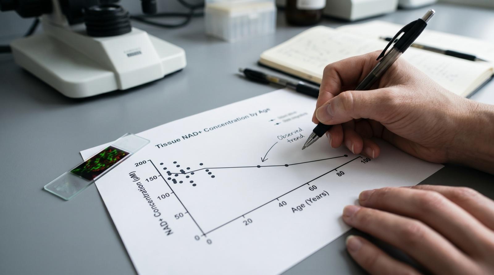
The acute scenario above is what happens in stroke, reperfusion injury, or a high dose of a chemotherapy drug. What happens in aging is the slow version. Decades of accumulated oxidative lesions, replication errors, and telomere-associated breaks keep PARP1 partially switched on all the time. The pool never crashes, but it never fully refills either. This chronic, low-grade drain is one of three converging mechanisms behind age-related NAD+ decline, alongside rising CD38 hydrolase activity and falling NAMPT salvage capacity.
PARP1 outcompetes sirtuins: the Km problem
NAD+ is consumed by three enzyme families — sirtuins, PARPs, and CD38/CD157 — and they do not draw from the pool on equal terms. The kinetics decide who gets fed when NAD+ is scarce.
Sirtuins have relatively high Michaelis constants for NAD+, mostly in the 100-300 μM range, and low maximal velocities. PARP1 has a Km roughly 5-fold lower and a much higher Vmax (Cantó et al. 2013, PMID 23357756; Houtkooper et al. 2010, PMID 20007326). Whole-cell NAD+ is usually reported at 200-500 μM, but free nuclear NAD+ — the fraction not bound to proteins — is lower, and genetically encoded sensors put it near the Km values of the signaling enzymes (Cambronne and Kraus 2020, PMID 32595066).
The practical consequence: when PARP1 is activated, it does not merely lower NAD+, it preferentially lowers the fraction available to sirtuins. Bai and colleagues showed the mirror image in 2011 (PMID 21459330). PARP1-knockout mice had higher NAD+ in brown adipose tissue and skeletal muscle, higher SIRT1 activity, more mitochondria, greater energy expenditure, and protection from diet-induced obesity. A pharmacological PARP inhibitor reproduced the NAD+ rise and SIRT1 activation in cells and in mice. The same logic, run in the opposite direction, was shown in worms: Mouchiroud et al. (2013, PMID 23870130) extended lifespan by either inhibiting PARP or supplementing NR, both acting through the NAD+/sirtuin axis and the mitochondrial unfolded protein response.
Two caveats keep this from becoming a simple “PARP1 bad, sirtuins good” narrative. First, Cantó's group notes that PARP2 has a Km and turnover similar to SIRT1, so PARP2 is unlikely to starve sirtuins the way PARP1 can. Second, the DBC1 data below make it clear that aging tissue does not have too much PARP1 activity — it has too little of the repair kind and, under acute stress, too much of the depleting kind. The companion piece on sirtuins and NAD+ covers the sirtuin side of this competition in depth.
| NAD+ consumer | Compartment | Trigger | Km for NAD+ | Products | Trend with age |
|---|---|---|---|---|---|
| PARP1 | Nucleus | DNA strand breaks, chromatin signals | ~20-60 μM | Poly-ADP-ribose + nicotinamide | Chronic low-grade activation; repair-mode activity falls (DBC1) |
| Sirtuins (SIRT1-7) | Nucleus, cytosol, mitochondria | Constitutive; NAD+-limited | ~100-300 μM | Deacylated protein + O-acetyl-ADP-ribose + nicotinamide | Activity falls as free NAD+ falls |
| CD38 / CD157 | Plasma membrane, endolysosome | Inflammation, senescence signals | Low micromolar | ADP-ribose, cyclic ADP-ribose + nicotinamide | Expression rises sharply (Camacho-Pereira et al. 2016, PMID 27304511) |
What is DBC1, and why is it called a brake on PARP1?
DBC1 — Deleted in Breast Cancer 1, also called CCAR2 — is one of the more abundant proteins in the mammalian nucleus and, for years, one of the least understood. Its name comes from a chromosomal deletion in some breast tumors, not from any established tumor-suppressor function. It was first placed in NAD+ biology in 2008, when two groups reported simultaneously in Nature that DBC1 binds SIRT1 and inhibits its deacetylase activity (Kim, Chen, and Lou 2008, PMID 18235501; Zhao et al. 2008, PMID 18235502). Escande and colleagues later showed that DBC1-knockout mice have higher hepatic SIRT1 activity and are protected from high-fat-diet liver steatosis (Escande et al. 2010, PMID 20071779).
The structural oddity that made DBC1 interesting is a domain that looks like a Nudix hydrolase — a family of enzymes that cleave nucleoside diphosphates — but lacks the catalytic residues to actually hydrolyze anything. Li and colleagues in David Sinclair's laboratory asked what a catalytically dead Nudix homology domain (NHD) might be for. Their 2017 Science paper (PMID 28336669) supplied the answer: it is an NAD+ binding pocket, and its occupancy controls which proteins DBC1 can touch.
The NAD+-DBC1-PARP1 switch: how aging turns DNA repair down
The core findings of Li et al. (2017) fit together as a single mechanism, so it is worth laying them out in order.
- DBC1 binds and inhibits PARP1. In vitro, purified DBC1 reduced PARP1 activity. In cells, knocking down DBC1 raised PAR levels both before and after exposure to paraquat, hydrogen peroxide, or etoposide, and increased expression of PARP1-regulated genes. Binding did not require PARP1 catalytic activity; a catalytically dead PARP1 mutant (E988K) still bound DBC1.
- NAD+ breaks the complex, and only NAD+ does. The PARP1-DBC1 interaction was dissolved by NAD+ in a concentration-dependent manner within the physiological range. At 200 μM, NMN, NR, adenosine, ATP, and ADP-ribose had no effect; 500 μM nicotinamide had no effect; 2 mM of the PARP inhibitor 3-aminobenzamide had no effect. The switch reads NAD+ specifically — not its precursors, not its breakdown products.
- The pocket is the NHD. Radiolabeled and biotin-labeled NAD+ bound DBC1 directly and could be competed off with unlabeled NAD+. Mutating a single predicted binding residue (Q391A) or deleting part of the NHD (residues 354-396) reduced NAD+ binding and made the PARP1-DBC1 complex insensitive to NAD+. Reintroducing wild-type DBC1 into knockdown cells re-suppressed PARP1; reintroducing the Q391A mutant did not.
- Releasing PARP1 improves repair. DBC1 knockdown lowered γH2AX (a marker of double-strand breaks), reduced DNA fragmentation in comet assays after paraquat, increased cell survival, and increased both non-homologous end joining and homologous recombination in a PARP1-dependent manner. NMN (500 μM) reduced γH2AX foci in paraquat-treated primary human fibroblasts.
- The switch flips with age in vivo. Livers of old mice (22-26 months) had lower NAD+, more DBC1-PARP1 complex, more γH2AX, lower PARP1 activity, and a blunted PARP1 response to new DNA damage than 6-month-old livers. DBC1-knockout mice aged 18-20 months had higher PARP1 activity than wild-type littermates.
- NMN reverses it in a week. Seven days of NMN at 500 mg/kg per day, given intraperitoneally, raised hepatic NAD+, dissociated the PARP1-DBC1 complex in both young and old mice, restored PARP1 activity in old mice (unless PARP1 was pharmacologically inhibited), and reduced γH2AX. In 23-26-month-old mice exposed to 7.5 Gy of gamma irradiation, NMN reduced DNA damage and protected white blood cell counts, lymphocytes, and hemoglobin — including when NMN was started after irradiation.
One brake, two enzymes: DBC1 on SIRT1 and PARP1
DBC1's inhibition of SIRT1 was known before its inhibition of PARP1, and the two interactions are regulated differently. In the Li et al. experiments, NAD+ disrupted the PARP1-DBC1 complex across the physiological range but left the SIRT1-DBC1 complex intact except at 500 μM. Other work shows the SIRT1-DBC1 interaction is controlled mainly by post-translational modification — DBC1 phosphorylation by ATM/ATR after DNA damage, for instance, tightens its grip on SIRT1 (Zannini et al. 2012, PMID 22735644).
The result is a single protein that coordinates the two largest nuclear NAD+ consumers and reads the cell's NAD+ status to do it. When NAD+ is high, DBC1 releases PARP1 and repair capacity is maximal. When NAD+ is low, DBC1 suppresses PARP1 — which, paradoxically, protects the remaining NAD+ pool for other uses at the cost of slower repair. Whether that trade-off is an adaptive program or a failure mode is a matter of interpretation, but the outcome in old animals is unambiguous: more unrepaired damage.
PARP1 capacity and lifespan: the Bürkle correlation
The DBC1 data sit inside a longer thread of evidence that DNA repair capacity, and PARP1 capacity specifically, tracks longevity. Grube and Bürkle (1992, PMID 1465394) measured maximally stimulated PARP activity in mononuclear leukocytes from 132 individuals across 13 mammalian species and found a strong positive correlation with species maximum lifespan (r = 0.84), with human cells showing about five times the activity of rat cells. Within species, PARP activity declined only weakly with donor age.
Muiras and colleagues extended this to humans (1998, PMID 9587069), comparing lymphoblastoid cell lines from 49 French centenarians against 51 controls aged 20-70. Maximal PARP activity was significantly higher in centenarian-derived cells (median 10,380 vs 9,035 cpm per million cells, P = 0.031), and specific activity per unit of PARP protein separated the groups even more clearly. Both studies are correlational and were done in stimulated, permeabilized cells, so they measure capacity rather than in vivo activity — but the direction is consistent with the DBC1 model: the organisms and individuals that live longest are the ones that can run PARP1 hardest when it counts.
Chronic PARP1 activation: what progeroid syndromes teach
Human diseases of defective DNA repair provide a natural experiment in what unrelenting PARP1 activation does to a cell. Three papers from the Bohr laboratory built the case:
- Xeroderma pigmentosum group A.Fang et al. (2014, Cell, PMID 24813611) found that XPA-deficient patient fibroblasts and Xpa-knockout mouse tissue accumulate unrepaired lesions that keep PARP1 chronically active, draining NAD+ to roughly half of control levels. The NAD+ deficit suppressed SIRT1, which impaired PGC-1α-dependent mitophagy, and damaged mitochondria piled up. NR, NMN, or PARP inhibition each rescued mitochondrial function. The site's research summary of the XPA paper walks through the cascade in detail.
- Cockayne syndrome. Scheibye-Knudsen et al. (2014, Cell Metabolism, PMID 25440059) showed the same PARP1 hyperactivation, NAD+ loss, and SIRT1 suppression in Cockayne syndrome models, rescued by NAD+ repletion or a high-fat (ketogenic) diet.
- Ataxia telangiectasia. Fang et al. (2016, Cell Metabolism, PMID 27732836) reported that NR extended lifespan and improved neuromuscular function in ATM-deficient worms and mice, acting through both mitophagy and DNA repair. This is the preclinical basis for the small human NR study in A-T patients discussed below.
These syndromes are the extreme end of a spectrum. Normal aging involves the same wiring — damage, PARP1 activation, NAD+ drain, sirtuin suppression, mitochondrial quality-control failure — at a fraction of the intensity. They are also the clearest human rationale for NAD+ repletion in neurodegeneration, where DNA damage accumulates in post-mitotic neurons; the Alzheimer's disease review covers PARP1 elevation in that context.
Acute collapse: PARP1, parthanatos, and energy failure
At the opposite extreme from slow aging is PARP1-dependent cell death, now called parthanatos. Yu et al. (2002, Science, PMID 12114629) showed that PARP1 hyperactivation causes the mitochondrial protein apoptosis-inducing factor (AIF) to translocate to the nucleus and trigger caspase-independent death. Andrabi et al. (2006, PMID 17116882) then identified the PAR polymer itself as the death signal that drives AIF release. Kolthur-Seetharam et al. (2006, PMID 16628003) tied this to sirtuin competition: SIRT1 activity limits PARP1-mediated AIF release, and NAD+ depletion by PARP1 removes that restraint.
The energy side is equally direct. PARP1 depletion of NAD+ starves glycolysis and the mitochondrial electron transport chain of their electron carrier; ATP falls; the cell dies necrotically. Pillai et al. (2005, PMID 16207712) showed this cascade in cardiomyocytes under pressure overload, where PARP1 activation reduced NAD+ and SIRT1 activity and caused cell death that PARP inhibition prevented. The same cascade explains why PARP1-knockout mice are resistant to cerebral ischemia (Eliasson et al. 1997, PMID 9334719) — and it is the reason acute NAD+ depletion is a different clinical problem from chronic aging decline. For the redox side of why NAD+ loss cripples ATP production, see NAD+ vs NADH.
Does NMN improve DNA repair? Grading the evidence
The honest answer has three layers, and the evidence tier drops at each one.
| Claim | Best evidence | Model | Tier (per /methodology) |
|---|---|---|---|
| NMN restores PARP1 activity and lowers DNA damage in aged tissue | Li et al. 2017 (PMID 28336669) | Mice (22-26 months), primary human fibroblasts | Preclinical |
| NAD+ repletion rescues mitophagy and function in DNA-repair-deficient disease | Fang 2014 (PMID 24813611); Scheibye-Knudsen 2014 (PMID 25440059); Fang 2016 (PMID 27732836) | Patient cells, worms, mice | Preclinical |
| NR improves ataxia scores in ataxia telangiectasia | Veenhuis et al. 2021 (PMID 34515380) | 24 patients, open-label, 4 months | Emerging |
| Nicotinamide reduces new non-melanoma skin cancers | Chen et al. 2015, NEJM (PMID 26488693) | 386 patients, phase 3 RCT, 12 months | Moderate |
| NMN or NR improves DNA repair capacity in healthy humans | No trial with DNA repair as a primary endpoint | — | Not graded (no human data) |
Layer one — animals and cells. Li et al. (2017) is the strongest mechanistic demonstration: NMN restored PARP1 activity, reduced γH2AX, improved survival after paraquat, and protected old mice from radiation, and it did so through a molecular switch that was mapped to a single residue. The dose, 500 mg/kg per day intraperitoneally, is far above anything used in human trials, and the endpoint was a DNA damage marker rather than a health outcome. It sits squarely in the preclinical tier.
Layer two — human proof of concept in rare disease. Veenhuis et al. (2021, PMID 34515380) gave NR to 24 patients with ataxia telangiectasia for four months in an open-label design. Ataxia scores improved during treatment and worsened after withdrawal, serum IgG rose in immunodeficient patients, and no adverse effects were reported. It is a small uncontrolled study in a genetic DNA-repair disorder — encouraging, and squarely emerging rather than moderate.
Layer three — the one moderate-tier human result. The ONTRAC trial (Chen et al. 2015, NEJM, PMID 26488693) randomized 386 people with a history of non-melanoma skin cancer to 500 mg nicotinamide twice daily or placebo for 12 months. New skin cancers were 23% lower in the nicotinamide group (P = 0.02). The proposed mechanism — from Surjana et al. (2013, PMID 23349012) — is that nicotinamide replenishes NAD+ in UV-stressed keratinocytes and enhances repair of UV-induced lesions, consistent with PARP1 needing substrate to function. Note this is nicotinamide, not NMN or NR, and the outcome is skin cancer incidence in a high-risk population, not a measure of repair kinetics. The broader comparison of NAM, NR, and NMN explains why the three precursors are not interchangeable.
PARP inhibitors and nicotinamide: two ways to change the equation
If PARP1 drains NAD+, why not simply inhibit it? Two reasons, beyond the DBC1 evidence that aging tissue needs more repair activity rather than less.
First, clinical PARP inhibitors are not gentle NAD+-sparing agents. Olaparib and the other approved clinical PARP inhibitors work in oncology by trapping PARP1 on DNA, converting single-strand lesions into replication-fork collapse and double-strand breaks that homologous-recombination-deficient tumors cannot repair (Murai et al. 2012, PMID 23118055). The cytotoxicity is the therapeutic mechanism. The metabolic PARP-inhibitor experiments in Bai et al. (2011) used research compounds in mice; they are proof of principle for the NAD+-sirtuin link, not a route to human longevity dosing.
Second, the cell already has a built-in PARP1 governor: nicotinamide, the product of every PARP1 reaction, is a feedback inhibitor of both PARPs and sirtuins at high-micromolar to millimolar concentrations. At physiological levels (tens of micromolar) it is instead the main substrate for NAD+ resynthesis through NAMPT. The skin cancer trial dose sits in the repletion range, not the inhibition range, and Li et al. found 500 μM nicotinamide did nothing to the DBC1-PARP1 complex. Nicotinamide's dual identity as PARP1 product, PARP1 inhibitor, and NAD+ precursor is one of the reasons the precursor field is more complicated than “raise NAD+” suggests — see the glossary entry on PARP and the DNA repair benefit page for the reference-level framing.
Where PARP1 gets its NAD+: local synthesis at chromatin
One refinement to the “PARP1 drains the pool” picture: the nucleus does not rely solely on diffusion from the cytosol. Ryu et al. (2018, Science, PMID 29748257) showed that NMNAT1, the nuclear enzyme that converts NMN to NAD+, is recruited to specific gene promoters where PARP1 is active and supplies NAD+ locally to sustain PARP1-dependent transcription. Cambronne and Kraus (2020, PMID 32595066) extended this into a general model of compartmentalized NAD+ pools, in which the nuclear pool is maintained partly independently of the cytosolic one.
The implication for supplementation is that the last step of NMN conversion — NMN to NAD+ — can happen right where PARP1 needs it, which is mechanistically tidy. It does not resolve the upstream question of how much oral NMN reaches the nucleus of an aging hepatocyte or neuron in the first place; that remains an open pharmacokinetic problem.
Open questions
- Does the DBC1 switch operate in human tissue? Li et al. showed it in human cells and mouse liver. No study has measured DBC1-PARP1 complex abundance in human biopsies across age or after NAD+ precursor supplementation.
- Which compartment matters? Chronic PARP1 drain and DBC1 inhibition are nuclear events. Blood NAD+ measurements do not report nuclear NAD+, and the available human assays cannot yet resolve compartments.
- Is more PARP1 always better? The Bürkle correlations suggest high repair-mode PARP1 capacity tracks longevity, but PARP1 also co-activates NF-κB and inflammatory transcription (Hassa and Hottiger 2002, PMID 12440774). The distinction between PARP1 at DNA breaks and PARP1 at promoters is not something current interventions can target selectively.
- Can NAD+ precursors protect against radiation or chemotherapy in humans? The irradiation protection in Li et al. (2017) is the most translatable finding in the paper and has not been tested clinically.
Bottom line
PARP1 is the enzyme that turns DNA damage into NAD+ loss. It uses NAD+ as its only substrate, spends one molecule per ADP-ribose unit, and builds polymers that are torn down within a minute — so sustained damage means sustained consumption. Under acute stress that can halve the pool in minutes and kill the cell through energy failure. Under chronic aging it is one of three drains, alongside CD38 and falling NAMPT, that lower the NAD+ available to sirtuins, which PARP1 outcompetes on kinetics.
The DBC1 discovery closed the loop. When NAD+ falls, DBC1 binds and inhibits PARP1, so aging tissue ends up with less repair capacity precisely when it has more damage to repair. Restoring NAD+ with NMN reopened that switch in old mice within a week — a preclinical result, but one mapped down to a single amino acid.
For humans, the evidence is proportionate: moderate for nicotinamide reducing skin cancer in high-risk patients, emerging for NR in ataxia telangiectasia, and absent for NMN or NR improving DNA repair in healthy adults. The mechanism is real; the human outcome data are still to come.
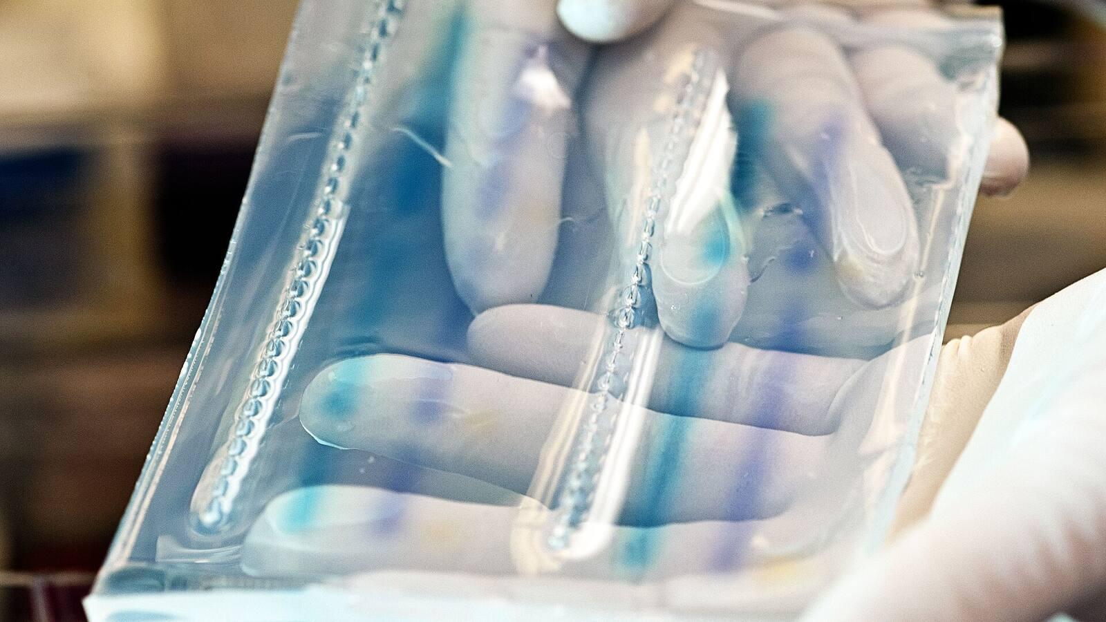
Cover image: “Agarose gel slab for DNA analysis, after the electrophoresis run” by the University of Michigan School of Natural Resources and Environment, via Wikimedia Commons, licensed under CC BY 2.0. Cropped and resized.
Frequently asked questions
- How does PARP1 use NAD+?
- PARP1 uses NAD+ as its only substrate. When it binds a DNA strand break, it cleaves the bond between nicotinamide and ADP-ribose in NAD+, releases the nicotinamide, and attaches the ADP-ribose unit to itself or to nearby proteins. It then repeats this reaction to build branched poly-ADP-ribose (PAR) chains of up to about 200 units. Every unit costs one NAD+ molecule, and the chains are degraded by PARG within about a minute, so an active PARP1 site cycles through hundreds of NAD+ molecules in a short time (Cantó et al. 2013, PMID 23357756; Gibson and Kraus 2012, PMID 22713970).
- Why does DNA damage lower NAD+?
- Because PARP1 activation is proportional to strand-break load and its catalytic rate is high. Under severe genotoxic stress, PARP1 activity can drop cellular NAD+ by 50-70% or more within minutes to an hour, which in turn starves ATP synthesis and can trigger necrotic cell death. Fibroblasts lacking PARP1 are protected from this NAD+ and ATP collapse (Ha and Snyder 1999, PMID 10570184; Berger 1985, PMID 3155867; Cantó et al. 2013, PMID 23357756). With aging, the damage load is lower but chronic, so the drain is slower and persistent rather than acute.
- What is DBC1 and how does it interact with PARP1?
- DBC1 (Deleted in Breast Cancer 1, also called CCAR2) is an abundant nuclear protein with a Nudix homology domain that binds NAD+ directly. Li et al. (2017, Science, PMID 28336669) showed that when NAD+ occupies this pocket, DBC1 cannot bind PARP1. When NAD+ falls, as it does with age, DBC1 binds and inhibits PARP1, reducing DNA repair capacity. In 22-month-old mice, DBC1-PARP1 complexes were elevated and PARP1 activity was lower; one week of NMN (500 mg/kg/day intraperitoneally) restored hepatic NAD+, dissociated the complex, restored PARP1 activity, and reduced the DNA damage marker gamma-H2AX. DBC1 also independently inhibits SIRT1 (Kim et al. 2008, PMID 18235501; Zhao et al. 2008, PMID 18235502).
- Does NMN improve DNA repair?
- In mice and in cultured human cells, yes: NMN restored PARP1 activity in aged mouse liver, reduced gamma-H2AX after paraquat exposure in primary human fibroblasts, and protected old mice from radiation-induced DNA damage and blood-count changes (Li et al. 2017, PMID 28336669). In humans, no randomized trial has measured DNA repair capacity as a primary endpoint after NMN or NR. The closest human evidence is indirect: high-dose nicotinamide (500 mg twice daily) reduced new non-melanoma skin cancers by 23% over 12 months in a phase 3 trial (Chen et al. 2015, NEJM, PMID 26488693), and an open-label NR study in 24 ataxia telangiectasia patients improved ataxia scores (Veenhuis et al. 2021, PMID 34515380). Under the NADFaq evidence grading at /methodology, NMN for DNA repair is preclinical; nicotinamide for UV-damage outcomes is moderate.
- Do PARP inhibitors raise NAD+ levels?
- In animal and cell studies, yes. PARP1 knockout mice and mice treated with a PARP inhibitor showed higher NAD+ in muscle and brown fat, more SIRT1 activity, greater mitochondrial content, and protection from diet-induced obesity (Bai et al. 2011, PMID 21459330). But clinical PARP inhibitors such as olaparib are oncology drugs that trap PARP1 on DNA and cause cytotoxic replication stress (Murai et al. 2012, PMID 23118055). They are not a longevity strategy, and the whole point of the DBC1 story is that healthy aging appears to need more PARP1 repair activity, not less. Do not take PARP inhibitors to preserve NAD+.
- Does nicotinamide inhibit PARP1?
- At high concentrations, yes. Nicotinamide (NAM) is the product released by every PARP1 reaction, and it feeds back to inhibit PARP1 and sirtuins in the high-micromolar to low-millimolar range. At physiological concentrations (tens of micromolar) it is the main precursor for NAD+ salvage via NAMPT, so it supports rather than suppresses PARP1 activity. This dual role is why nicotinamide dosing matters and why the 500 mg twice-daily dose in the skin cancer trial is best interpreted as NAD+ repletion for repair, not PARP1 blockade (Surjana et al. 2013, PMID 23349012; Chen et al. 2015, PMID 26488693).